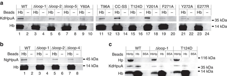
Figure 5. The affect of HpuA mutations on pull down by Hb.
(a) Coomassie-stained gel showing pull-down results of Hb and control (−) beads with wild type (WT) and mutated KdHpuA protein including deletion mutants, single point mutants and mutation of the Cys–Cys motif (residues 119–120) to Ser–Ser (CC–SS). (b) Pull-down results with Hb and control beads pulling down NgHpuA wild type and loop deletions. (c) Binding of KdHpuA wild type and mutants to HbHp beads, a similar loss of binding is observed as compared with Hb beads alone. The positions of molecular weight markers are shown to the right of each gel slice. Input samples from these pull downs are shown in Supplementary Fig. 8.

