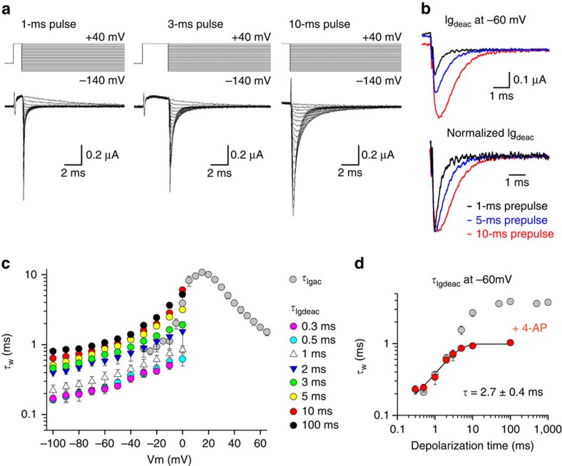
Figure 4. Kv3.1b relaxes from a pre-active state in the presence of 4-AP.
(a) Igdeac currents of Kv3.1b in the presence of 1 mM 4-aminopyridine (4-AP) with different activating pulse durations at +40 mV. (b) Top panel, superposition of Igdeac at −60 mV on a 1-ms (black), 5-ms (blue) and 10-ms (red) long depolarization to +40 mV. Bottom panel, superposition of the same Igdeac currents after normalization. Note the gradual deceleration in Igdeac decay as the activating pulses are prolonged. (c) Igdeac kinetics (τIgdeac) as a function of depolarization time in the presence of 1 mM 4-AP, displayed as mean±s.e.m. (n=5). Even in the presence of 1 mM 4-AP there was a noticeable deceleration in τIgdeac that developed relatively quickly. For example, compare τIideac on a 0.3-ms (pink) and a 3-ms (green) activating pulse at+40 mV. (d) Weighted τIgdeac at −60 mV as a function of the activating pulse duration in both the absence (grey) and presence of 1 mM 4-AP (red), shown as mean±s.e.m. (n=5). τIgdeac was slowed down in presence of 4-AP, a deceleration that developed over a similar time window as the fast slowing process (τf) in control conditions.

