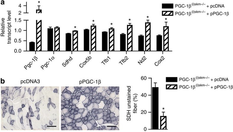
Figure 6. Rescue of mitochondrial defects in skeletal muscles of PGC-1β(i)skm−/− mice.
(a) Relative transcript levels, determined by RT-qPCR, of PGC-1β and α, Sdhd, Cox5b, Tfb1, Tfb2, Nd2 and Cox2 in gastrocnemius muscle of 26-week-old PGC-1β(i)skm−/− mice, 7 days after electroporation with an empty vector (pcDNA3) or a plasmid encoding PGC-1β (pPGC-1β; n=4). (b) Histological staining of tibialis anterior from 18-week-old PGC-1β(i)skm−/− mice, 7 days after electroporation with an empty vector (pcDNA3, left panel) or a plasmid encoding PGC-1β (right panel), for succinate dehydrogenase activity, and quantification of unstained fibers (n=4). Scale bar, 100 μm. Data are represented as mean±s.e.m. *P<0.05, Student's t-test.

