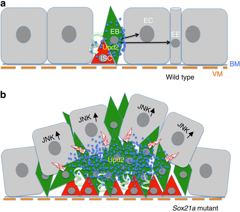
Figure 9. Model of Sox21a tumour initiation and progression.
(a) Schematic representation of a wild-type intestinal epithelium. Intestinal stem cells (ISCs) are localized basally close to the basement membrane (BM) and visceral muscles (VMs). ISCs self-renew and differentiate to generate differentiating progenitors, the enteroblasts (EBs), which will then further differentiate into either enterocytes (ECs) or enteroendocrine (EE) cells. Progenitors express Upd2 (blue dots) stimulating basal level ISC turnover. (b) The Sox21a mutation blocks the differentiation of EBs to ECs or EE cells, resulting in the accumulation of EBs. Clustered EBs create a centre with high Upd2 level that simulates ISCs division, generating more differentiation-defective EBs. EB tumour cells eliminate flanking ECs by delamination probably under the action of ROS and mechanical pressure. Elimination of flanking ECs, a process requiring JNK signalling activation, further provides space and mitogens allowing tumour progression.

