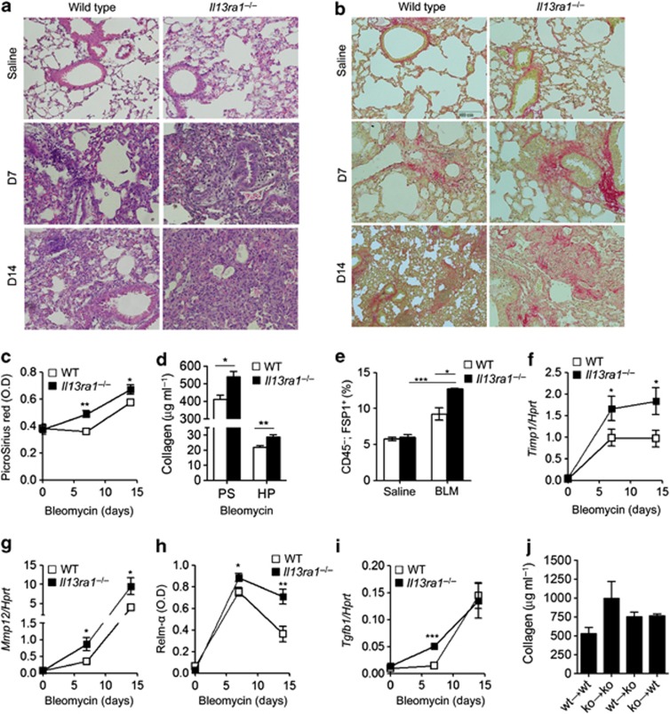
Figure 4.
Increased bleomycin-induced pathology in Il13ra1−/− mice. Representative photomicrographs of H&E (a) and Picro-Sirius red staining (b), as well as quantitative assessment of soluble collagen (c) and hydroxyproline content (d) in saline- and bleomycin-treated lungs at day 7 (D7) and 14 (D14) are shown. (e) Flow cytometric analysis of CD45+/FSP1+ fibroblasts in saline- and bleomycin (BLM)-treated wild-type and Il13ra1−/− mice. In j, soluble collagen levels in bone marrow chimeric mice at day 14 after bleomycin-treatment are shown. The expression of Timp1 (f), Mmp12 (g), and Tgfb1 (i) following saline or BLM treatment was assessed by qPCR analysis and normalized to the house keeping gene hypoxanthine-guanine phosphoribosyltransferase (Hprt). (h) The secretion of Relm-α was assessed by ELISA; n=3, *P<0.05, **P<0.01, ***P<0.001. H&E, hematoxylin and eosin; HP, Hydroxyproline; PS, Picro Sirius red.

