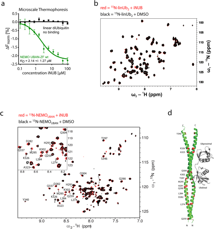
Figure 4. iNUB binds and alters the conformation of the NEMOUBAN.
(a) Microscale Thermophoresis (MST) assays to determine binding of iNUB to NEMOUBAN or linUb2. iNUB binds to Myc-NEMOUBAN-ZF-StrepTagII (242–350) with a KD of 2.14 μM. (b) 1H,15N HSQC spectra of 50 μM 15N-labeled linUb2 in the absence (black) and presence of 1 mM iNUB (red). (c) NMR analysis of the NEMOUBAN-iNUB interaction. 1H,15N TROSY NMR spectrum of 117 μM 15N-labeled NEMOUBAN (258–350) with DMSO (black) or 870 μM iNUB (red). Amide signals that are affected upon iNUB addition are indicated. Asterisks indicate unassigned NMR signals that are affected by iNUB. (d) Mapping of residues affected upon iNUB addition onto the NEMOUBAN crystal structure (PDB 2ZVO).

