
Figure 5. Percent distribution of coral mucus C assimilation into bacterial, sponge, coral, and algal phospholipid fatty acids (PLFAs).
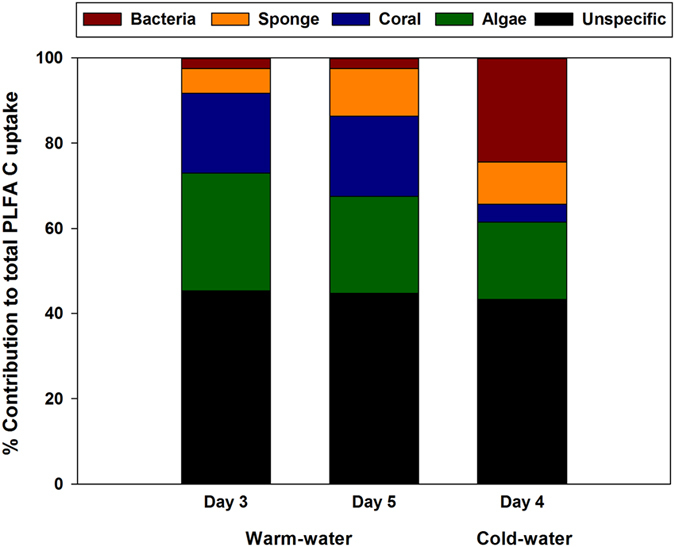
Data shown for warm-water sponge M. fistulifera after 3 and 5 days exposure to labeled coral mucus, and for the cold-water sponge H. coriacea after 4 days exposure to labeled coral mucus*. *Note that the cold-water PLFA data are from three sponge specimens maintained in a single experimental chamber during the labeling procedure.