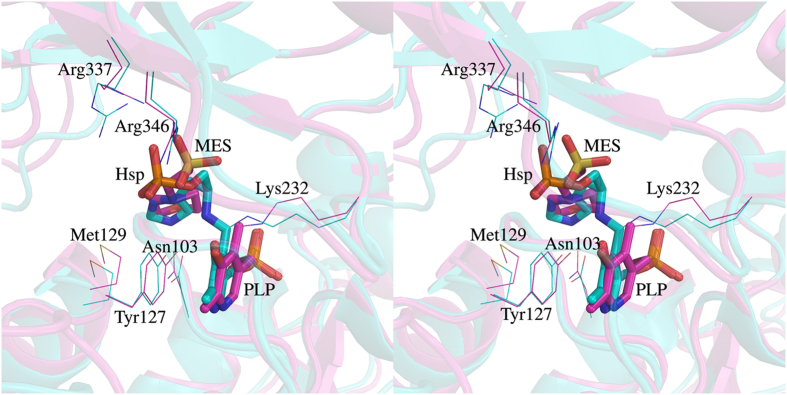
Figure 4. Comparison of the active site regions of Mtb and E. coli HspATs.
Structural differences between Mtb (magenta) and E. coli (cyan; P. D. B. ID: 1FG3) HspATs are presented in stereo mode. Both the structures were superimposed with 1.6 Å rmsd (over 639 Cα atom pairs). The active site residues in the both the structures are mostly conserved and MES in mHspAT binds in a manner similar to Hsp in eHspAT. The morphiline-ring of MES and the imidazole ring of Hsp overlap and their respective sulphate and phosphate groups are also positioned similarly. The PLP binding in both cases is almost identical. The labeled residues are with respect to mHspAT.

