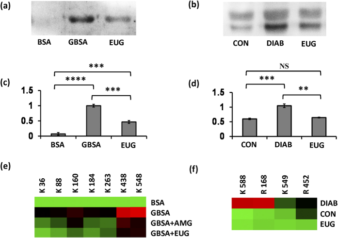
Figure 9. Proteomic analysis of in vitro and in vivo samples for AGE formation.
Western blot using anti-AGE antibody & blot density analysis of in vitro BSA-AGE assay samples (a,c) and in vivo plasma samples (b,d) for probing AGE formation. One way ANOVA followed by unpaired t-test suggested significant differences between data at p < 0.01 (indicated as ‘**’), p < 0.001 (indicated as ‘***’) and p < 0.0001 (indicated as ‘****’). NS represents non-significant difference in data. Heat map showing extent of AGE induced modifications on specific lysine and arginine residues in (e) in vitro BSA-AGE assay and (f) plasma protein, identified by LC-MSE. Heat map generated using Multi Experiment Viewer (MEV) software. (GBSA, glycated BSA; AMG, aminoguanidine; EUG, eugenol-treated sample; CON, control healthy mice; DIAB, STZ control plasma).

