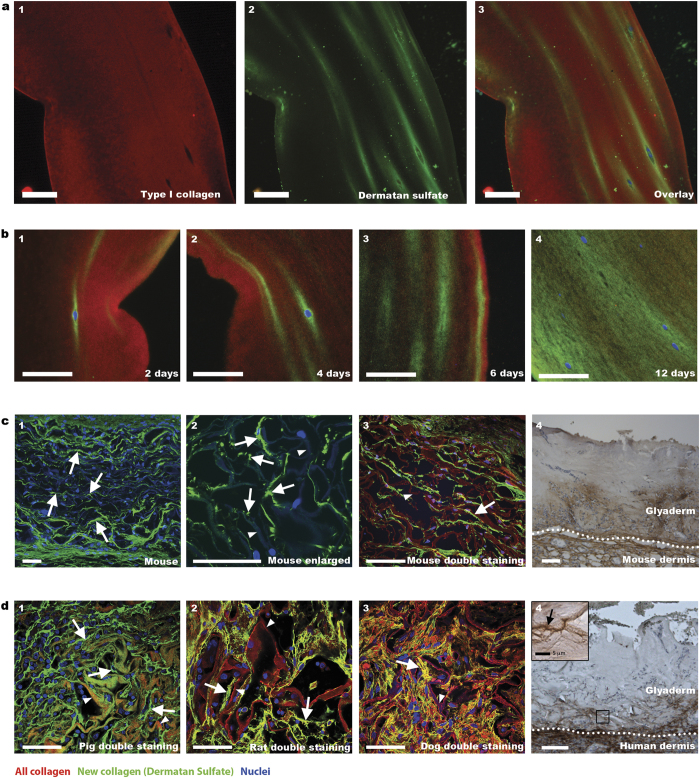
Figure 2. Detection of newly synthesized collagen fibrils in cellularized/implanted collagenous biomaterials.
(a) Collagen gel cultured for 6 days with human fibroblasts. Newly deposited collagen is indicated by green dermatan sulfate staining (a2,a3), whereas all collagen is indicated by red type I collagen staining (a1,a3). (b) Location of newly deposited collagen in collagen gels cultured in time with fibroblasts/keratinocytes. Note increase of new collagen over time (b1-4). (c) Newly deposited collagen fibrils (arrows) in a collagen scaffold (arrowhead), two weeks after subcutaneous implantation in mice (c1-3) (for clarity, autofluorescence of background collagen was enhanced). For Glyaderm® (acellular human dermis), new collagen is indicated by brown staining (c4). (d) Newly deposited collagen fibrils (arrow) in various species (d1-4) after implantation of a collagen scaffold (arrowhead) in pig (1 month) (d1), Integra® (arrow head) in rat (1 week) (d2), Integra® (arrow head) in dog (4 weeks) (d3), and Glyaderm® in human (inset shows fibrillar structure) (d4). Scale bars are 50 μm unless indicated otherwise.

