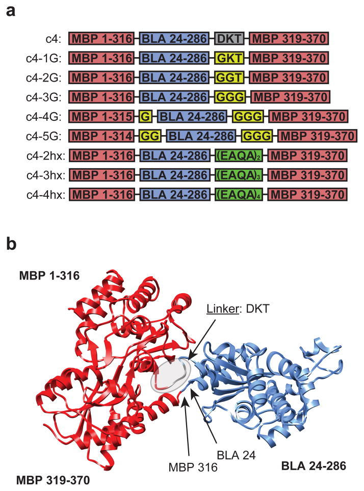
Figure 2.
Sequences and structural models of c4 and c4 variants. (a) Graphical illustration of the sequences of c4 and c4 variants indicating the MBP domain (red), BLA domain (blue), engineered glycine linker (yellow), and EAQA linker (green). (b) Structural model of the c4 fusion protein comprised of maltose binding protein (MBP; red) and TEM-1 β-lactamase (BLA; blue) domains with the DKT linker and fusion sites indicated by arrows.

