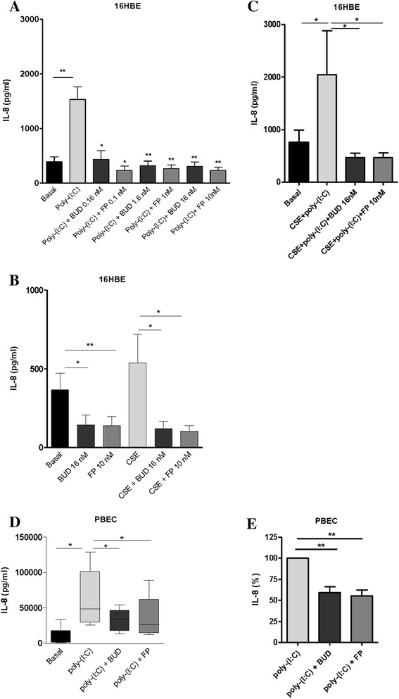
Fig. 1.

Effect of equivalent concentrations of budesonide (BUD) and fluticasone propionate (FP) on cigarette smoke extract (CSE) and/or poly-(I:C)-induced IL-8 (CXCL8) release in 16HBE cells and primary bronchial epithelial cells (PBECs). 16HBE cells and PBECs were seeded in duplicates, grown to confluence for 3–4 days, serum-deprived (16HBE) or placed on basal medium with transferrin and insulin (PBECs) overnight, pre-treated with or without 0.16-16 nM BUD or 0.1−10 nM FP for 2 hours and stimulated with 5 % CSE and/or 12.5 μg poly-(I:C) for 24 hours. IL-8 was measured in cell-free supernatants. a Mean ± SEM IL-8 levels (pg/ml) in 16HBE cells at baseline and upon stimulation with poly-(I:C) in the presence and absence of different concentrations of BUD and FP (n = 4). b Mean ± SEM IL-8 levels (pg/ml) in 16HBE cells at baseline and upon stimulation cells with 5 % CSE in the presence and absence of 16 nM BUD and 10 nM FP (n = 4). c Mean ± SEM IL-8 levels (pg/ml) in 16HBE cells upon combined stimulation with CSE + poly-(I:C) in the presence and absence of 16 nM BUD and 10 nM FP (n = 4). PBECs were exposed to 12.5 μg/ml poly-(I:C) for 24 hours and absolute IL-8 levels (pg/ml, min to max) (d) or values normalized to the absence of BUD or FP (percentage, mean ± SEM) (e) are shown (n = 6). * = p < 0.05 and ** = p < 0.01 between the indicated values or between the absence and presence of BUD or FP as determined by the Student’s t-test (assumed normal distribution) (a-c, e) or the Wilcoxon-signed rank test (d)
