Fig. 1.

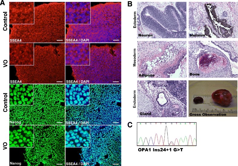
Characterization of iPSCs obtained from patient’s skin cells carrying an OPA1 mutation. a Control and VO-iPSCs (patient-derived) were stained with antibodies against pluripotency cell surface markers SSEA4 (red) and transcriptional factor Nanog (green). DAPI staining of nuclei is shown in blue. Scale bars = 20 μM. b Hematoxylin and eosin staining of VO-iPSC-derived teratomas shows cell lineages derived from three germ layers, the ectoderm (neuron, skin), mesoderm (blood vessel, bone), and endoderm (gland). A gross observation of dissected teratomas is also shown (left, kidney capsule; right, testis). c A representative sequencing trace that confirmed the presence of an OPA1 mutation (NM_015560.2(OPA1):c.2496 + 1G > T) in a VO-iPSC line
