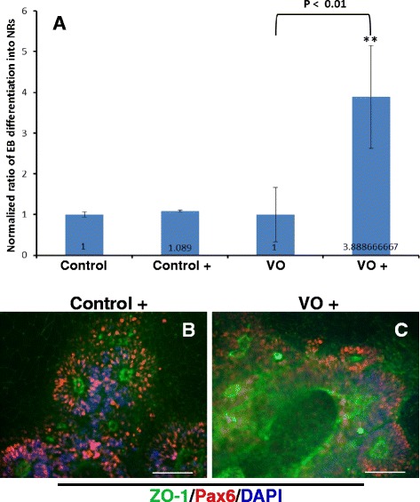
Fig. 5.

Control and VO-iPSCs were cultured in hESC medium containing 10 % FBS with and without 10 % NIM. Neural rosettes (NR) were stained with ZO-1 (green) and Pax6 (red) antibodies. In panel a, with (Control+ and V0+) and without (control & V0)10 % NIM Embryoid bodies (EBs) that exhibited a typical “rosette” structure were counted. Student’s t-test was used to analyze the data. All samples were normalized to the control values. *p < 0.01. Scale bars = 20 μM. Representative images are shown in panels b and c
