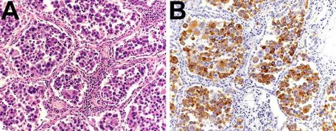
Figure 3.
Immunohistochemistry. (A) Tumor cells are discohesive, markedly pleomorphic, and fill almost completely the alveolar spaces of the lung parenchyma (hematoxylin and eosin (HE) staining). (B) Tumor cells show a diffuse cytoplasmic and focal nuclear immunostaining with S100 (a melanocyte marker).

