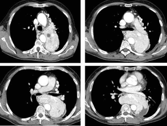
Figure 1.

Axial post-contrast computed tomography (CT) images at four different levels of the chest (shown here at the mediastinum window level), disclosed a large left-posterior mediastinal lesion characterized by many intralesional and surrounding vessels, strictly adherent to the mediastinal pleura and the left lung, and very close to the esophagus (medially dislocated by the mass).
