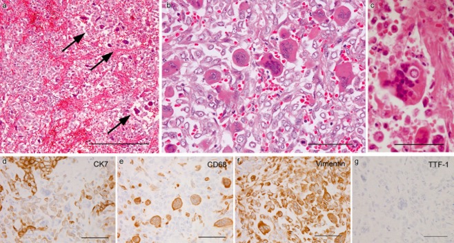
Figure 2.
Metastases containing osteoclast-like giant cells (OCGCs). (a) OCGCs chiefly distributed in the central portion (arrows) of gastric metastasis (bar = 200 μm). (b) Poorly cohesive carcinoma cells intermingled with numerous OCGCs in gallbladder metastasis (bar = 50 μm). (c) Giant cell phagocytizing an isolated carcinoma cell in rectal metastasis (bar = 50 μm). (d–g) Immunohistochemical findings in the same area of a small intestinal metastasis. Cytokeratin 7 (CK7) expression is strong in nested carcinoma cells, but weak in poorly cohesive carcinoma cells admixtured with CK7-negative OCGCs (d). CD68 staining only in OCGCs and a few histiocytes (e). Vimentin expression in OCGCs and many isolated carcinoma cells (f). No expression of thyroid transcription factor 1 in metastatic lesion (G) (d–g, bars = 50 μm)

