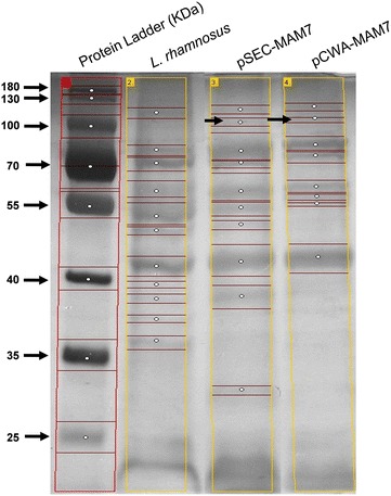
Fig. 2.

SDS-PAGE electrophoresis from recombinants L. rhamnsus strains. The figure shows the protein profile from L. rhamnosus pSEC-MAM-7 (pSEC-MAM7), L. rhamnosus pCWA-MAM-7 (pCWA-MAM7) and L. rhamnosus wild type (L. rhamnosus). Black arrows show a weakly band of approximately 100 kDa in recombinants strains. Bands of each lane were detected using GelAnalyzer and each white circle indicates a band
