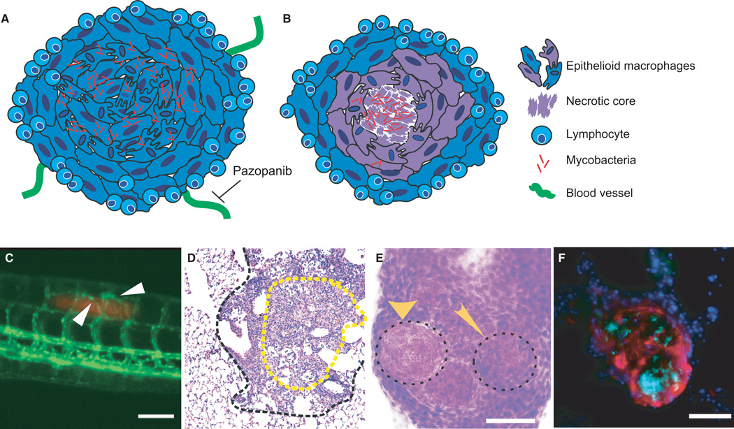
Fig. 1. Heterogeneous granuloma progression leads to morphologically and phenotypically distinct granuloma subtypes.
(A) Schematic of a normoxic, non-necrotic granuloma. A non-necrotic granuloma is comprised of a core of infected and uninfected epithelioid macrophages, with a cuff of lymphocytes. Vasculature extends toward the granuloma, and can be inhibited by the VEGFR inhibitor pazopanib. (B) Schematic of a necrotic, hypoxic granuloma. A mycobacteria-containing central core of necrotic cell debris is surrounded by layers of epithelioid macrophages circumscribed by a lymphocytic cuff. This granuloma is characterized by hypoxia (represented in purple) within the core. (C) Vascularization of early mycobacterial granulomas. Angiogenesis induced by granuloma formation in a 6-day post infection zebrafish larva infected with fluorescent M. marinum (red). Vasculature (flk1:gfp transgenic line) in green. White arrows point to blood vessels sprouting toward the site of infection. (D) H&E stain of a typical mouse granuloma. Outlined in a black dashed line is a granuloma from a C57Bl/6 mouse 25 weeks post-infection with Mycobacterium tuberculosis. The central region of epithelioid macrophages (eosin-rich region in yellow dotted line) is surrounded by a lymphocyte-rich cuff, with little evidence of a necrotic core or hypoxia. (E) H&E staining of adult zebrafish granuloma shows distinct necrotic and non-necrotic granulomas. A cryosectioned adult zebrafish infected with M. marinum for 2 weeks. The wide arrow marks a necrotic granuloma and the narrow arrow shows a non-necrotic granuloma, indicated by absence and presence of hematoxylin staining within the central core. (F) Necrotic granulomas are hypoxic in adult zebrafish. Section of an adult zebrafish infected with M. marinum 2 weeks post-infection, stained for pimonidazole (red), DAPI (blue), and M.marinum (cyan). Hypoxia, indicated by pimonidazole staining, is seen in and around a central mycobacteria-containing necrotic core. Scale bars in all panels are 100 µm.

