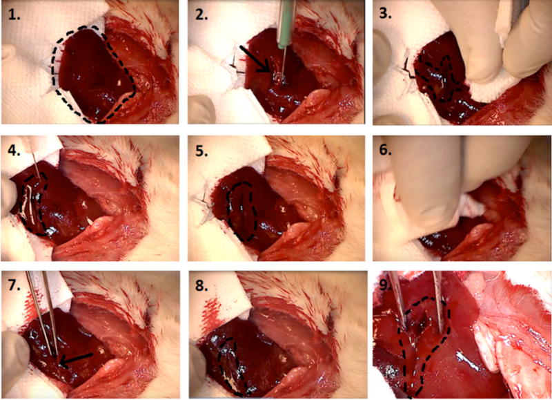
Figure 4.

Liver incision model: step-by-step procedure in applying SB50. 1. Exposed left lobe of liver preincision (right cranial, left caudal). 2. Immediately after lateral incision by scalpel (arrow points at incision). 3. Profuse bleeding prior to application of SB50 gel (indicated by area marked by dotted line). 4. Application of SB50 gel after wiping cut with sterile gauze. Gel is localized to the area within the dotted line on image, right above the incision. 5. SB50 gel left on cut for 2 min. 6. SB50 gel is wiped away at the end of a 2 min waiting period. 7. Cut (indicated by arrow) is perturbed by probing with sterile tweezers. 8. No bleeding is observed even after excessive perturbation. Incision site is marked by dotted line. 9. Zoomed in image of cut showing no bleeding during site perturbation.
