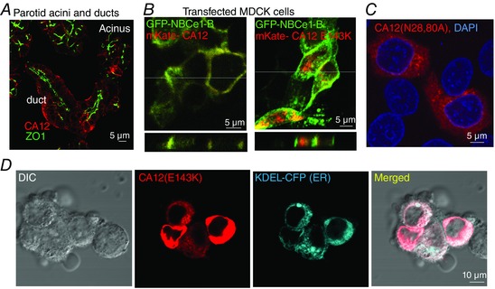
Figure 1. Localization of native and expressed CA12 and mutants .

A, localization of CA12 (red) and ZO1 (green) in dispersed parotid acini and ducts. B, x–y (upper panels) and z scan (lower panels, taken at the white line in the upper panels) images of MDCK cells transfected with mKate‐CA12 (red) and GFP‐NBCe1‐B (green) (left image) or with mKate‐CA12(E143K) and GFP‐NBCe1‐B (right panel). C, HEK cells transfected with CA12(N28,80A) (red) and stained for DAPI (blue). D, co‐localization of CA12(E143K) (red) and the ER marker KDEL‐CFP (blue) in transfected in HEK cells.
