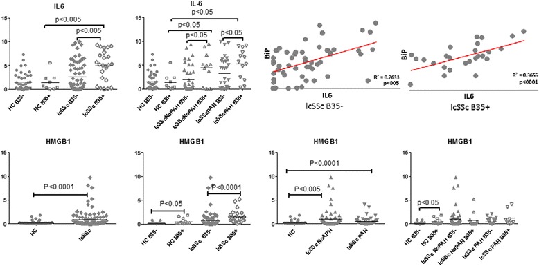
Fig. 3.

IL-6 and HMGB1 are elevated in B35-positive subjects. PBMCs were isolated from HC (n = 49), lcSSc (n = 97, NoPAH n = 53 and PAH n = 44) and grouped according to the presence of the HLA-B*35 allele: HC B35+ (n = 9), HC B35- (n = 40); lcSSc B35+ (n =26), lcSSc B35- (n =71); lcSSc NoPAH B35+ (n =14), lcSSc NoPAH B35- (n = 39), lcSSc PAH B35+ (n = 12) and lcSSc PAH B35- (n = 32). mRNA levels of IL-6 (top panel) and HMGB1 (bottom panel) were determined by qPCR. Expression of the housekeeping genes β-actin, GADPH, and 18S served as internal positive controls. Data are expressed as the fold-change normalized to mRNA expression in a single HC sample. Each data point represents a single subject; horizontal lines show the mean. Right top panel depicts linear regression analysis of the relationship between expression of BiP and IL-6 in PBMCs from lcSSc B35-negative and B35-positive patients. IL-6 interleukin-6, HMGB1 high-mobility group protein B1, PBMCs, peripheral blood mononuclear cells, HC healthy controls, lcSSc limited cutaneous systemic sclerosis, PAH pulmonary arterial hypertension
