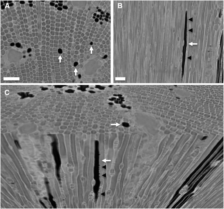
Figure 3.
Imaging from microCT scans demonstrating the presence of isolated, embolized tracheids (white arrows) in the stem xylem of P. pinaster for a transverse slice (A), a longitudinal slice (B), and a three-dimensional rendering showing transverse and longitudinal views together (C). Gas in bordered pit chambers connecting an embolized and a functional tracheid can be observed in longitudinal slices (black arrowheads). Isolated tracheids were not connected to other resolvable gas-filled spaces in the xylem of intact plants. Bars = 50 μm.

