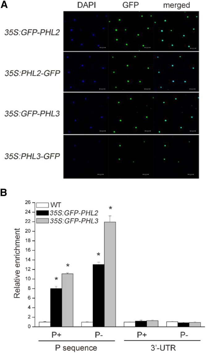
Figure 5.
PHL2 and PHL3 are localized in the nucleus and bind to the P sequence in vivo. A, Subcellular localization of PHL2 and PHL3. The expression constructs as indicated on the left were infiltrated into the leaves N. benthamiana and GFP fluorescence was observed 2 d after infiltration. The nuclei were stained by DAPI. B, ChIP-PCR analysis of the in vivo binding of the GFP-PHL2 or GFP-PHL3 to the P sequence. Chromatins from the wild type and the transgenic plants expressing GFP-PHL2 or GFP-PHL3 were immunoprecipitated with GFP-Trap. The levels of enrichment of the precipitated DNA fragments were quantified by qPCR assay. Values are means ± se of three replicates. Asterisks indicate a significant difference from the wild type under the same growth conditions (Student’s t test, P < 0.05).

