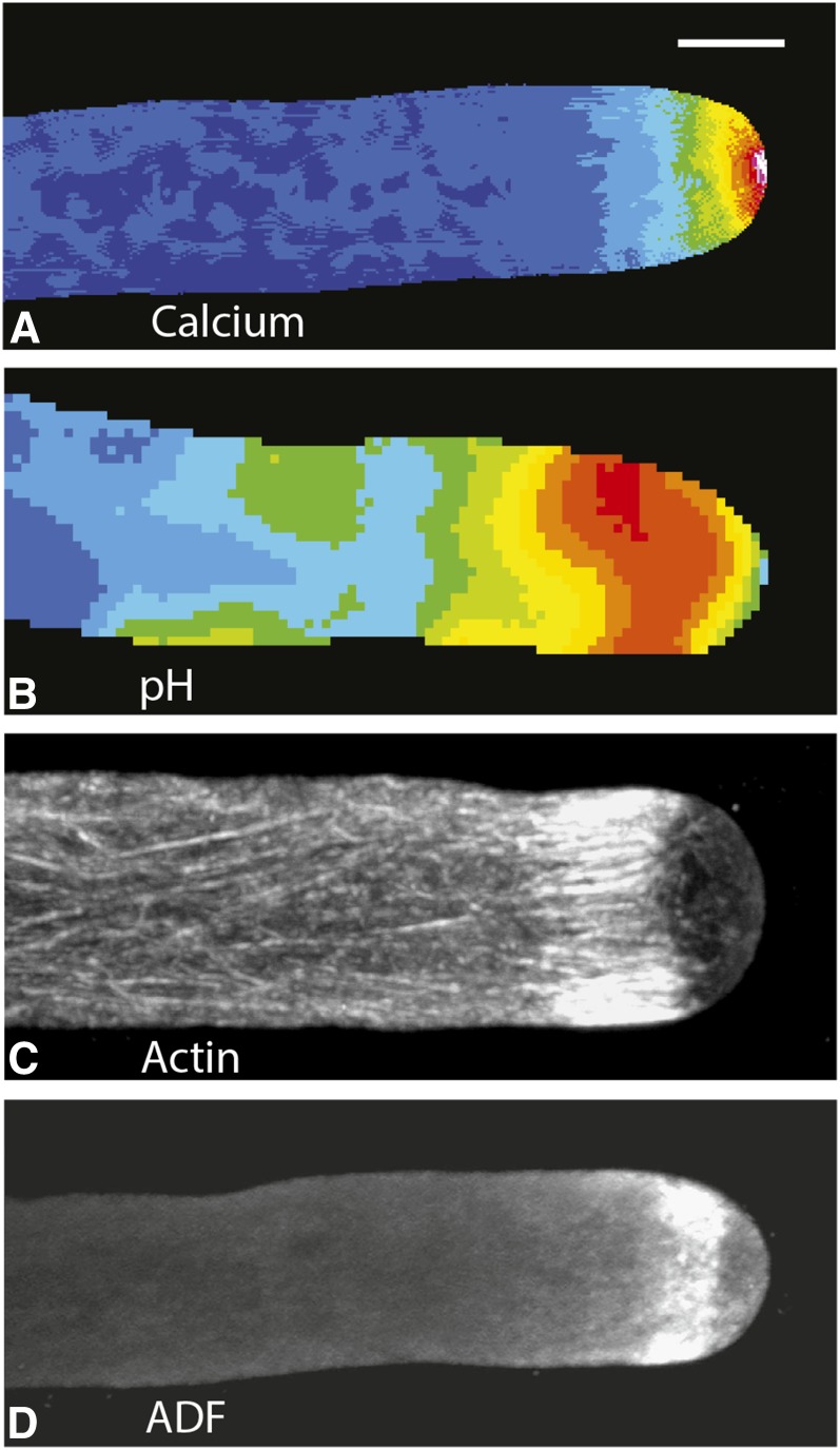
Figure 1.
Lily pollen tubes, which have been treated with different reporters or stains, show the distribution of free calcium (A), pH (B), F-actin (C), and the actin-binding protein ADF (D). A, Microinjection of the calcium-sensitive dye Fura-2-dextran allows one to image the distribution of free calcium in a living lily pollen tube. Ratiometric analysis reveals that the calcium is maximal at the extreme apex, where the concentrations are 1 to 10 µm. Back from the tip, the calcium concentration drops sharply, reaching the basal level of 0.15 µm within 15 to 20 µm. From Rounds et al. (2011). B, In this instance, the living pollen tube has been microinjected with BCECF-dextran, a pH-sensitive dye. The ratiometric image reveals a slightly acidic region at the extreme apex followed by a prominent alkaline band (orange/red; pH 7.5), starting a few micrometers behind the tip and extending rearward for an additional 10 to 20 µm. Thereafter, the pH approaches neutrality. From Rounds et al. (2011). C, In this image, the lily pollen tube has been preserved through rapid freeze fixation, followed by rehydration and staining with an anti-actin antibody. The resulting confocal fluorescence image reveals the striking cortical actin fringe that begins a few micrometers back from the tip and extends rearward for 5 to 10 µm. The MFs in the fringe are organized as a palisade in which the individual elements are aligned parallel to the long axis of the pollen tube. Behind the fringe, the MFs are also longitudinally oriented, but they appear to be much less dense than in the fringe. From Lovy-Wheeler et al. (2005). D, In this example, a lily pollen tube, which has been preserved through rapid freeze fixation and rehydration, has been stained with an antibody to lily ADF1. The stained region starts a few micrometers back from the apex and extends rearward 3 to 5 µm. By comparison with C, it is obvious that ADF colocalizes with the actin fringe, perhaps especially the forward edge of the fringe. Also note that ADF (D) and the actin fringe (C) colocalize with the alkaline band (B). From Lovy-Wheeler et al. (2006). Bar, 10 μm.

