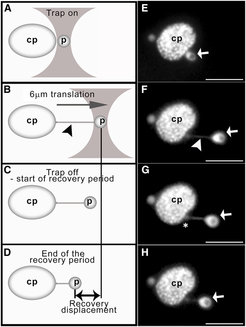
Figure 1.
Optical trapping and movement of peroxisomes away from chloroplasts in tobacco leaf epidermal cells. Schematic representations of the trapping procedure (A–D) and corresponding micrographs (E–H) are shown. Upon turning the trap on (A and E) and moving the stage 6 μm at a set speed (B and F; referred to as the translation period), the trapped peroxisome (p; white arrows) is pulled away from the cp and a peroxule (arrowheads) is formed. Upon turning the trap off (C and G), the peroxisome recoils backs toward its original position next to the chloroplast (D and H). Peroxisome displacement during the recovery period (referred to as recovery displacement) is measured (double-headed arrow). The asterisk denotes the tip of the peroxule. Bars = 6 μm.

