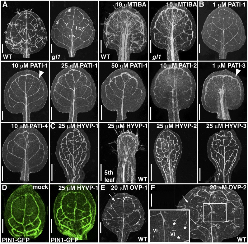
Figure 1.
Compound-induced vein pattern defects. A to C, First rosette cleared leaves of 12-d-old gl1 seedlings (unless otherwise noted) from compound-grown plants. A, gl1 has similar compound responsiveness to 10 µm TIBA as wild type (WT). T, Trichomes; mv, midvein; al, apical loop; sl, second loop; lv, lateral vein; hov, higher order veins. B, PATI-induced phenotypes. Arrowhead indicates thick marginal ring. C, HYVP (25 µm)-induced vein patterns to show extensive venation network. Note elongated, wide petiole and reduced lamina of fifth leaf from HYVP-1-treated plants. D, Confocal images of PIN1-GFP-labeled incipient vascular cells in developing leaves shows that the extensive vein branching in 25 µm HYVP-1-treated leaves occurs at an early stage. E and F, First rosette cleared leaves of 12-d-old wild-type seedlings grown on media containing 20 µm OVP-1 (E) or 20 µm OVP-2 (F). Open loops are indicated with arrows. Boxed region of OVP-2 is at higher magnification (inset). VI, Vascular islands (short stretches of unconnected vein). Vascular cells have irregular morphology, bifurcated vein endings, and delayed differentiation (asterisks). Tracheary elements appear as white lines in cleared leaves viewed under dark-field microscopy. Bars, 500 µm (A–C, E, and F), or 100 µm (D).

