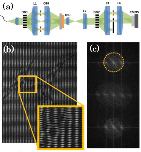
Fig. 1.

(a) Optical schematic for structured illumination diffraction phase microscopy (SI-DPM) system. (b) Raw interferogram taken of endothelial progenitor cells shows the structured illumination pattern overlayed on the carrier spatial frequency. (c) Fourier transform of raw interference pattern is shown with region of frequency-space to be filtered and DC centered outlined by dashed yellow circle.
