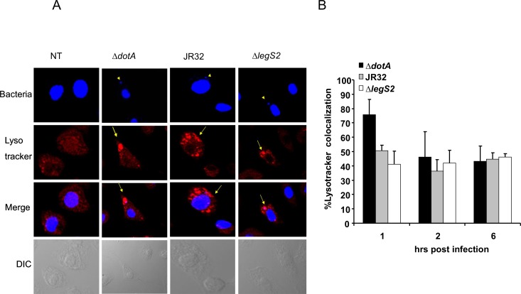
Fig 4. Replication of L. pneumophila legS2 mutant is not due to defective phagosome-lysosome fusion.
(A) Images of wild-type macrophages not infected (NT) or infected with the type IV secretion mutant dotA, wild-type L. pneumophila, JR32, and the legS2 mutant. The first panel presents staining with DAPI, with arrow heads pointing to the bacteria. The second panel shows staining with the lyso tracker. The third panel depicts merged images, with bacteria colocalized with the lysosomal marker. B) The percent of bacteria colocalized with the lyso tracker was scored in 100 infected cells from 3 independent coverslips. The data represents the mean + SD of n = 3.

