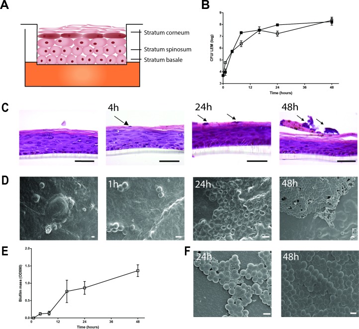
Fig 1. Biofilm formation by S. aureus LUH14616 on LEMs and PS surfaces.
(A) Schematic representation of LEM. (B) Bacterial counts were performed on LEM exposed to LUH14616 for various intervals. Adherent bacteria are represented by open symbols and non-adherent/loosely adherent bacteria by closed symbols. Results are displayed as the mean and SD of four experiments. (C) Haematoxilin and eosine staining of LEMs at various intervals after inoculation with LUH14616. Arrows indicate microcolonies, scale bars = 50 μm. (D) Cryo scanning electron microscopy of LEMs colonized with LUH14616 for various intervals. Photographs are representative for three different keratinocyte donors. (E) Biofilm formation by LUH14616 on PS in IMDM medium. Results are the mean and SEM of three experiments. (F) Cryo scanning electron microscopy of S. aureus LUH14616 biofilms formed on PS at 24 and 48 hrs after adherence to wells. Scale bars = 1 μm.

