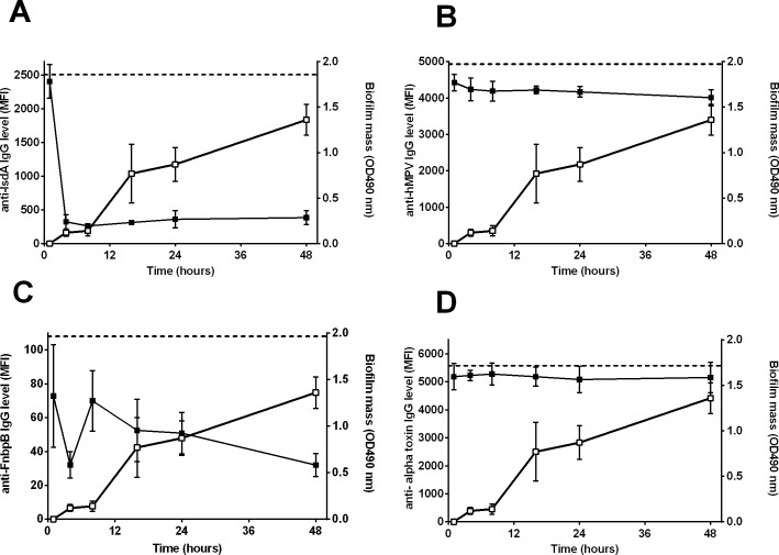
Fig 2. Detection of S. aureus proteins during biofilm formation of LUH14616 on PS.
Closed symbols indicate the mean fluorescence intensity (MFI, left Y-axis), reflecting the level of remaining non-bound IgG directed against specific proteins after incubation of PHG with the biofilms, while open symbols indicate biofilm mass (OD490 nm, right Y-axis). Both are plotted against the time of biofilm growth (hrs). Results are shown for (A) IsdA, (B) control protein of human metapneumovirus (hMPV), (C) FnbpB and (D) alpha toxin. Dashed horizontal lines indicate the average MFI of sterile controls. Symbols and error bars indicate mean and SD of four experiments.

