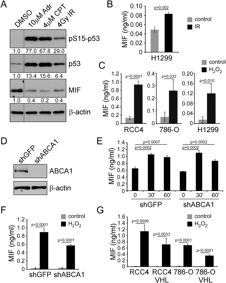
Fig 2. DNA damaging agents induce MIF secretion.
A) Western blot of RCC4 cells following 4 hours of treatment with 10 μM adriamycin, 4 μM camptothecin, or 4 Gy IR probed with phosphoserine 15 p53, total p53, MIF, and β-actin antibodies. B) MIF ELISA analysis for H1299 cells irradiated and tested at the one hour time point. C) MIF ELISA on RCC4, 786-O, and H1299 cell conditioned media after stimulation with 10 mM H2O2 for 2 hours. D) Western blot of RCC4 cell lysates with ABCA1 knockdown by shRNA compared to control shGFP probed with ABCA1 and β-actin antibodies. E) Secretion of MIF measured by ELISA from shABCA1 and shGFP RCC4 cells at 30 minutes and 60 minutes after 4 Gy IR. F) Secretion of MIF measured by ELISA from shABCA1 and shGFP RCC4 cells at 60 minutes after exposure to 10 mM H2O2. G) Secretion of MIF measured by ELISA from RCC4 and 786-O cells with or without VHL resonstitution at 60 minutes after exposure to 4 Gy IR.

