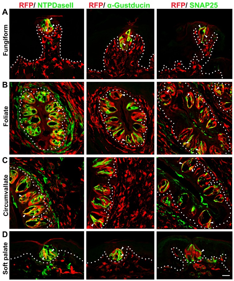
Fig 4. Dermo1-Cre labeled all three types (I, II, III) of taste bud cells in adult mice.
RFP+ signals were co-localized with markers for specific taste cell types (white arrowheads), i.e., NTPDaseII for type I cells, α-Gustducin for type II cells and SNAP25 for type III cells in the lingual (A, B, C) and palatal (D) taste buds. White dotted lines demarcate the epithelium from underlying connective tissue. Scale bar: 20 μm for all images (single plane laser-scanning confocal).

