Fig. 3.
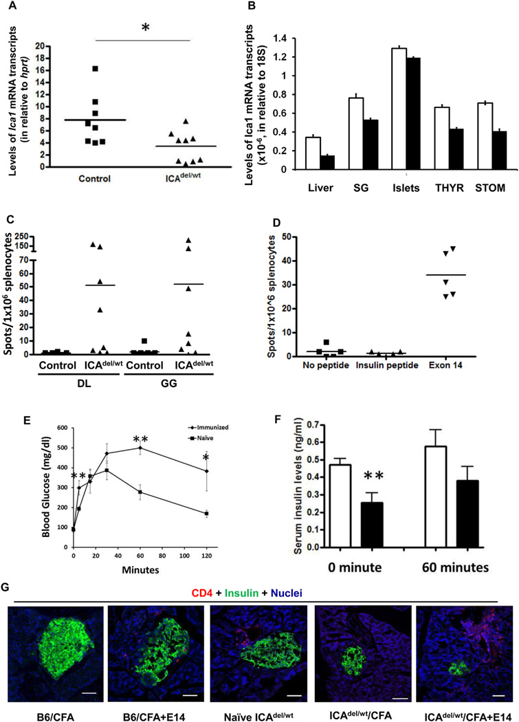
Ica1 haploid deficient mice (ICA69del/wt) develop anti-islet autoimmunity. A. RT-qPCR analysis of Ica1 mRNA transcript expression in CD45+ cell-depleted, thymic stromal cells of ICA69del/wt mice. Filled squares, wild-type littermate controls; filled triangles, ICA69del/wt thymic stroma samples. *p < 0.05. B. TaqMan RT-qPCR analysis of levels of Ica1 mRNA transcription in various extrathymic organs of ICAdel/wt mice. Total RNA samples were isolated from the liver, salivary glands (SG), islets, thyroid glands (THYR) and stomach (STOM) of 12-week old ICAdel/wt (n = 3, filled bar) and wild type control mice (n = 3, open bar). Absolute numbers of Ica1 mRNA transcripts were calculated and normalized to those of 18S. C. ELISPOT of IFNγ-producing cells in the spleens of naïve ICA69del/wt mice. Splenocytes were stimulated with ICA69 C-terminal peptides GG and DL (listed in Fig. 1A). Filled squares, wild type littermate controls; filled triangles, ICA69del/wt mice. D. Development of anti-ICA69 autoimminity in B6.ICA69del/wt mice immunized with E14. ELISPOT of IFNγ-secreting splenocytes harvested from ICA69del/wt mice, using E14 as stimulants. An H-2b restricted, insulin peptide (LWMRFLPL) was used as control. E. Intraperitoneal glucose tolerance test of ICA69del/wt mice immunized with exon 14 (n = 6), in comparison to non-immunized naïve ICA69del/wt mice (n = 6). Data are presented as mean ± SEM. *p < 0.05; **p < 0.01. F. Serum insulin levels in naïve (open bars, n = 7), or exon 14-immunized (filled bars, n = 6) mice. Sera were collected from ICA69del/wt mice either after overnight fasting (0 min), or 60 min after i.p. injection of a bolus of 2 g/kg of d-glucose. Data are presented as mean ± SEM. **p < 0.01. G. Immunohistochemistry of lymphocytic infiltration into the pancreata of E14-immunized ICA69del/wt mice. From left to right are representative cryosections of pancreata harvested from: B6 mice immunized with CFA, B6 mice immunized with CFA + E14 (16-weeks post immunization, only detectable in 2 out of 10 animals; not detectable in 4-weeks post treatment), naïve ICA69del/wt mice (24 weeks old, only detectable in 10% animals), CFA-immunized ICA69del/wt mice (16-weeks post treatment, detectable in 2 out 10 animals) and E14-immunized ICA69del/wt mice (16-weeks post treatment, detectable in majorities of the immunized animals) mice were stained with anti-CD4 (red) antibodies, and counter stained with anti-insulin (green) antibody. White arrows, infiltrating immune cells.

