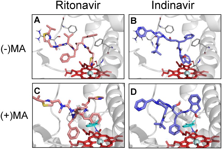
Fig 7. Docking simulations of Type II PIs in the absence and presence of methamphetamine.
Structures of (A) Ritonavir and (B) Indinavir with their preferred binding sites pointing towards the heme moiety of CYP3A4in the absence of methamphetamine. Docking simulations of (C) Ritonavir and (D) Indinavir with heme moiety of CYP3A4 in the presence of methamphetamine. R-methamphetamine in binding mode 1 was shown in cyan sticks. Docking was performed as described in Materials and Methods.

