Figure 1.
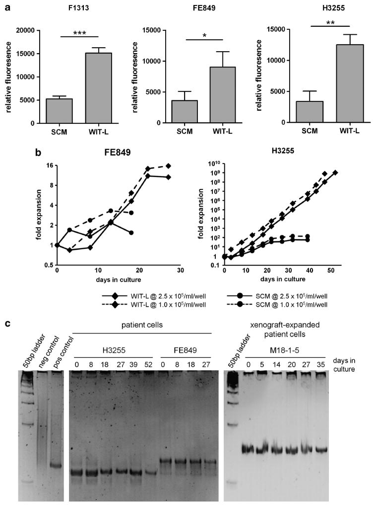
Serum-free WIT-L medium supports robust growth of primary T-ALL blasts in vitro. In vitro growth of primary T-ALL cells. (a) Resazurin reduction assay. T-lymphoblasts obtained directly from patient biopsy material were seeded in 24-well dishes at 2.5 × 10⁵ viable cells per well on confluent monolayers of irradiated (50 Gy) MS5-DL1 feeders in 1 ml of either SCM or WIT-L medium. Four to six days later, cell growth was measured by resazurin reduction assay (Cell Titer Blue, Promega, Madison, WI, USA) with subtraction of values obtained from feeder-only control wells. Error bars indicate s.d. of assays performed in triplicate. Data depicted are representative of 2–4 experimental replicates. *P<0.05; **P<0.01; ***P<0.001 (Student’s t-test). (b) Growth curves. Patient T-lymphoblasts were seeded in 24-well dishes at 1.0 ×10⁵ or 2.5 ×10⁵ cells per well on irradiated MS5-DL1 monolayers in 1 ml of either SCM or WIT-L medium. Cultures were passaged every 4–6 days by reseeding at the initial density onto fresh feeders. Carry-over of feeders between passages was minimized by filtering manually disaggregated cell suspensions through 48 μm nylon mesh. Viable cell yields were counted at each passage (Vi-Cell, Coulter, Brea, CA, USA). (c) TCRγ clonality assay by heteroduplex analysis. Genomic DNA from patient T-ALL samples cultured in WIT-L medium on MS5-DL1 feeders was PCR amplified using consensus TCRγ primers. Resulting PCR products were heated at 94 °C for 5 min, transferred to ice for 1 h, then run on a 12% non-denaturing polyacrylamide gel and visualized by ethidium bromide staining. Positive control is a biopsy-proven T-cell non-Hodgkin lymphoma; negative control is a reactive lymph node.

