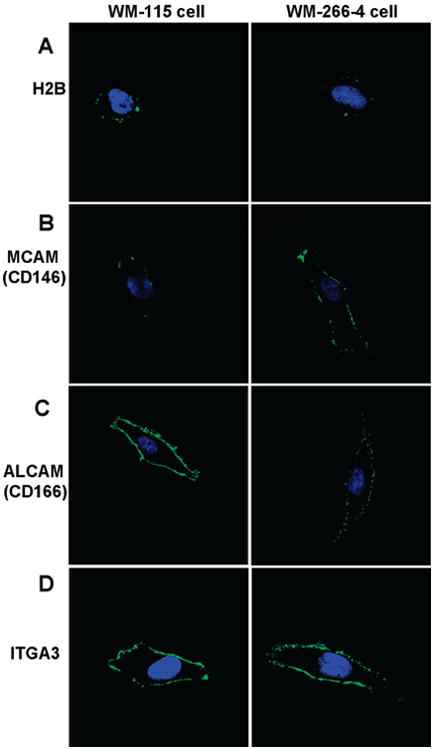
Figure 5.

Immunocytochemical detection of cell surface proteins. The left and right panels represent the staining of WM-115 and WM-266-4 cells, respectively. Cells were stained with primary mAbs to (A) H2B, (B) MCAM, (C) ALCAM, and (D) ITGA3 followed by staining with Alexa Fluor 488-labeled secondary antibody (green). Cells were also stained with DAPI (blue) to identify nuclei. Images of WM-115 and WM-266-4 cells stained with same primary mAbs were captured with identical parameters to compare the staining of the two cell lines.
