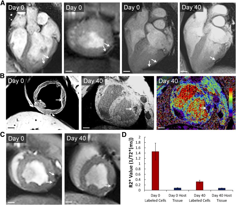
Figure 3.
In vivo detection of ferumoxytol-labeled human embryonic stem cell-derived cardiac progenitor cells (hESC-CPCs) in pig hearts by magnetic resonance (MR) imaging. (A–C): Day 0 and day 40, in vivo T2*-weighted MR images from three porcine hearts showing locations of ferumoxytol-labeled (arrows) hESC-CPCs; 4 × 10⁷ hESC-CPCs were injected. Scale bars = 1 cm. Labeling was done at day 0 of differentiation with 300 μg/ml of ferumoxytol. (D): R2* values (ms−1) measured from porcine host myocardium and labeled hESC-CPC injections at day 0 and day 40 (n = 3, mean ± SEM). R2* values were calculated from T2*-weighted MR images.

