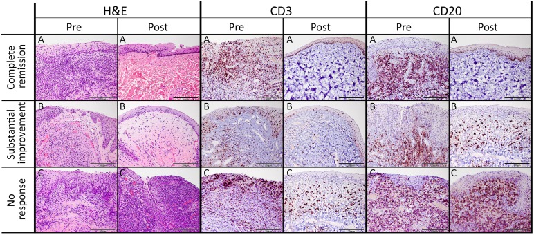
Figure 4.
Histologic and immunohistochemical evaluation of the feline chronic gingivostomatitis oral mucosa. Histomorphology of sections from all patients pretreatment was consistent with severe lymphoplasmacytic and neutrophilic stomatitis accompanied by epithelial hypertrophy and multifocal ulcerations ([A–C], H&E column). Pretreatment CD3+ T cells were present within the epithelium and submucosa, and CD20+ B cells were mainly present within the subepithelial stroma. Sections obtain from a cat with complete clinical remission ([A], Post columns) had histology consistent with normal mucosa. Histomorphology of sections from a substantial responder ([B], Post columns) was consistent with mild lymphoplasmacytic stomatitis accompanied by mild epithelial hyperplasia and superficial stromal edema. After treatment, moderate numbers of CD3+ T cells were observed within the epithelium and stroma in sections form partial responders, and moderate numbers of CD20+ T cells were located in the subepithelial stroma. Histomorphology of sections obtained from a nonresponder cat after treatment ([C], Post columns) was consistent with severe chronic lymphoplasmacytic and neutrophilic ulcerative stomatitis, which was histologically similar to treatment. Distribution of CD3+ and CD20+ cells was similar to that observed before treatment. Scale bar = 200 μm. Abbreviations: H&E, hematoxylin and eosin; post, after treatment; pre, before treatment.

