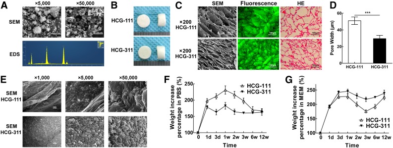
Figure 3.
Characteristics and property of HCG-111 and HCG-311 scaffolds. (A): SEM and EDS microanalysis for HA nanoparticles. Nanohydroxyapatite (nHA) showed a rod-like nanostructures (approximately 100 nm × 30 nm) at higher magnification (scale bars = 500 nm). However, at lower magnification (scale bars = 5 μm), nHA agglomerations were rougher lumps, approximately 1–2 μm. EDS spectra were obtained with the beam focused on the nHA particles. The predominant components were carbon, oxygen, phosphorus, and calcium (carbon detected by the conductive tapes). (B): Photographs showing the synthesized scaffolds were 8 mm in diameter × 5 mm thick. (C): SEM images, fluorescence observation, and H&E staining in the cross-sections showing the inner scaffold network connection and pore structure. Scale bars = 100 μm. (D): The measured pore width showed that HCG-111 had larger pores than HCG-311. Data are shown as mean ± SD (n = 16, ***, p < .001). (E): SEM images at different magnifications showed the HCG-311 pore wall had rougher structures. From left to right in sequence, scale bar = 10 μm, 1 μm, and 100 nm. (F): Absorption characteristics of scaffolds soaked in phosphate-buffered saline (PBS) as a function of time. HCG-111 had increased more in weight within 12 weeks. Data are presented as mean ± SD (n = 3). (G): Absorption capacity of scaffolds soaked in α-MEM (10% FBS) was greater with more protein than that for scaffolds soaked in PBS, stratified by time. HCG-311 uptake resulted in a greater weight within 12 weeks. Data are presented as mean ± SD (n = 3). Abbreviations: d, day; EDS, energy dispersive spectroscopy; HCG, nanohydroxyapatite/chitosan/gelatin; HCG-111, HCG scaffold with 1 wt/vol% nHA; HCG-311, HCG scaffold with 3 wt/vol% nHA; HE, H&E; MEM, α-modified minimum essential medium; PBS, phosphate-buffered saline; SEM, scanning electron microscopy; w, week.

