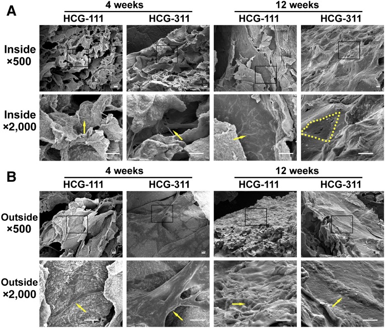
Figure 4.
Scanning electron microscope (SEM) images revealing the inside and outside appearance of in vitro-cultured tissues. (A): Inside the HCG-111 and HCG-311 scaffolds, the cells communicated and an intercellular bridge had formed (yellow arrow) at 4 weeks. Cells and membrane had blocked the pores (yellow dots outline inside pore margin) on HCG-311 or spread out along with the pore wall (yellow arrow) on HCG-111 at 12 weeks. (B): Outside appearance of cell adherence and proliferation at 4 weeks (yellow arrows) and membrane formation at 12 weeks (yellow arrows) on HCG-111 and HCG-311. Higher magnification images (bottom) show an expanded view of the selected areas (black rectangles) in the top images. Scale bars = 10 μm. Abbreviations: HCG-111, nanohydroxyapatite/chitosan/gelatin scaffold with 1 wt/vol% nanohydroxyapatite; HCG-311, nanohydroxyapatite/chitosan/gelatin scaffold with 3 wt/vol% nanohydroxyapatite.

