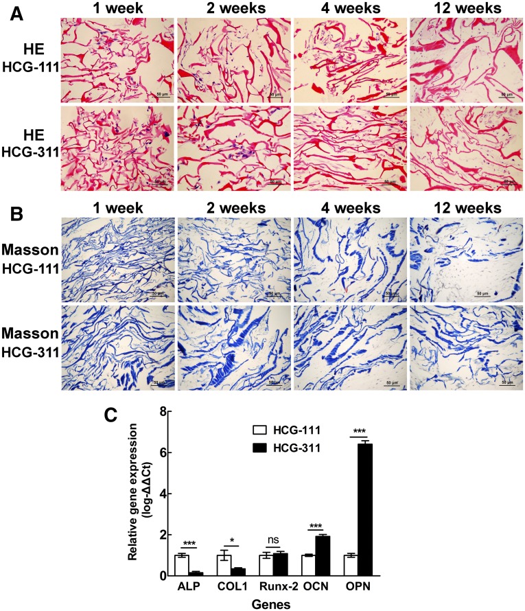
Figure 5.
Histologic and quantitative real-time polymerase chain reaction (qRT-PCR) assessments of tissues cultured in vitro. Samples of human induced pluripotent stem cell (hiPSC)/nanohydroxyapatite/chitosan/gelatin (HCG) complexes were collected and sectioned for histologic staining at 1, 2, 4, and 12 weeks. (A): Sections were stained with H&E staining. The cells adhered and proliferated well on both HCG-111 and HCG-311 scaffolds. Dense extracellular matrix had formed at 12 weeks. Scale bars = 50 μm. (B): Sections were stained with Masson trichrome staining. Collagen stained in blue. Scale bars = 50 μm. (C): Bone-associated gene (ALP, Col1, Runx-2, OCN, and OPN) expression in hiPSCs on HCG-111 and HCG-311 scaffolds were analyzed by qRT-PCR at 4 weeks. The 2−ΔΔCt method was used to analyze the results, where Ct values on HCG-311 were normalized to human 18S and further to the Ct values of the control HCG-111 samples. OCN and OPN expression in hiPSCs was significantly higher on HCG-311 scaffolds than on HCG-111 scaffolds (p < .001). Data are presented as mean ± SD (n = 3); *, p < .05; ***, p < .001. Abbreviations: HCG-111, nanohydroxyapatite/chitosan/gelatin scaffold with 1 wt/vol% nanohydroxyapatite; HCG-311, nanohydroxyapatite/chitosan/gelatin scaffold with 3 wt/vol% nanohydroxyapatite; ns, no statistically significant difference (p > .05).

