Figure 1.

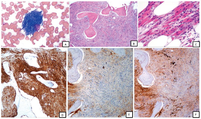
Case 1 (A-F): A, bone marrow touch imprint showing histiocytes with numerous crystalline inclusions within the cytoplasm, Giemsa-Wright stain, x1000; B-C, low and high power magnifications of bone marrow biopsy; marrow space demonstrates extensive spindle cell proliferation composed of polygonal-shaped histiocytes with crystalline cytoplasmic inclusions, H & E stain, x100 and x400; D, By immunohistochemistry, these histiocytes were positive for CD163 (x100, shown); CD68 (KP-1), SMA (focal) and IgG and negative for desmin, myogenin, EBER, melanoma cocktail, S-100, IgM, IgA (not shown); E and F, Staining for kappa and lambda immunoglobulin light chains highlight sparse plasma cells that show bright monotypic lambda staining; the spindle cells show weak blush of lambda and kappa (equivocal)
