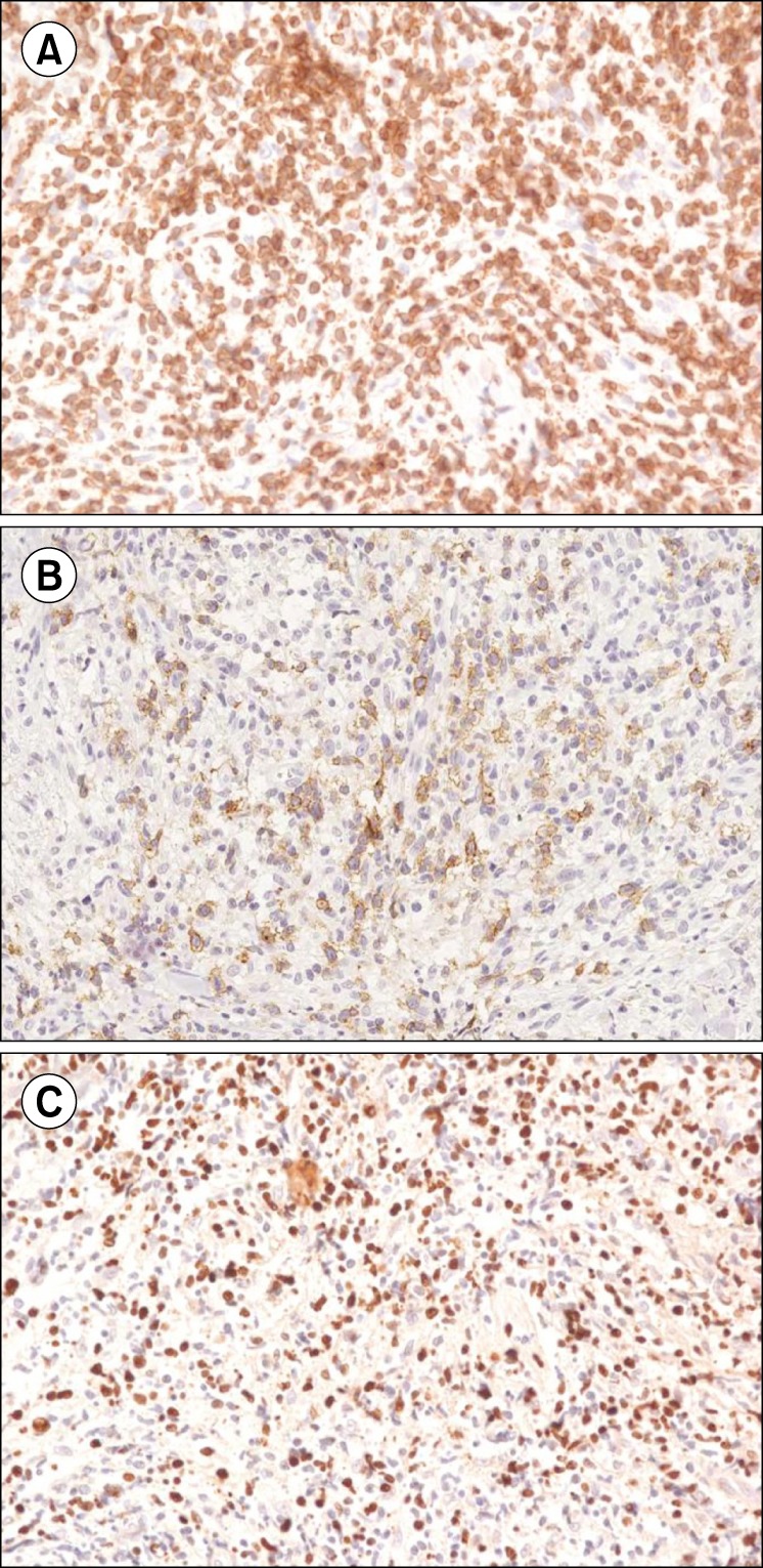
Fig. 2. Pathology images of the skin biopsy showing extranodal NK/T-cell lymphoma (ENKL). (A) Positive staining for CD3 in lymphoma cells (×400 magnification). (B) Positive staining for CD30 in lymphoma cells (×400 magnification). (C) In situ hybridization of Epstein-Barr virus, showing positive cells (×400 magnification).