Figure 2.
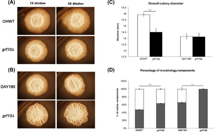
The grf10Δ mutant in two genetic backgrounds exhibits filamentation and colony defects. Cells (0.08 OD600) were spotted onto Spider medium; photos of the macroscopic colonies were taken on day 10: (A) strains OHWT and TF021 (SN152 background), (B) strains DAY185 and RAC117 (BWP17 background). (C) Quantification of the overall colony diameter (mm) at day 10. (D) Distribution of the overall colony diameter by percentage into the central region (dark bar) and peripheral region (white bar). All measurements were averaged from six biological samples with two technical replicates (n = 12). Student's t-test was calculated using Excel. *** p-value < 0.0001.

