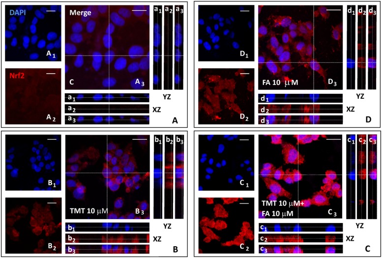
FIGURE 3.
Ferulic acid induces Nrf2 activation and translocation into the nucleus. (A–D) Representative images from four independent immunofluorescence experiments in which we performed a double-labeling with DAPI (A1–D1) and an anti-Nrf2 antibody (A2–D2). Merged images are shown in (A3–D3). XZ and YZ cross-sections in the boxes (referred to the dashed lines) from the confocal Z-stack acquisitions illustrate cytosolic or nuclear fluorescence signal(s) (XZ and YZ boxes: a1–d1 refer to DAPI staining; a2–d2: refer to Nrf2 fluorescence; a3–d3: Merge). In cells treated with TMT, a strong Nrf2 activation was detected which, however, remains mainly confined in the cytoplasm (b1–b3 in B). In TMT + FA treated cells, there was a further cytoplasmic enhancement of Nrf2 expression compared to TMT alone which also translocates into the nucleus as indicated by Z-stack acquisitions (c1–c3 in C). FA administration (10 μM) induced an endogenous antioxidant response leading to a strong increase in Nrf2 expression in the cytosol (d1–d3 in D) compared to Control (a1-a3 in A). Scale bar: 20 μm. For further information see text.

