FIGURE 5.
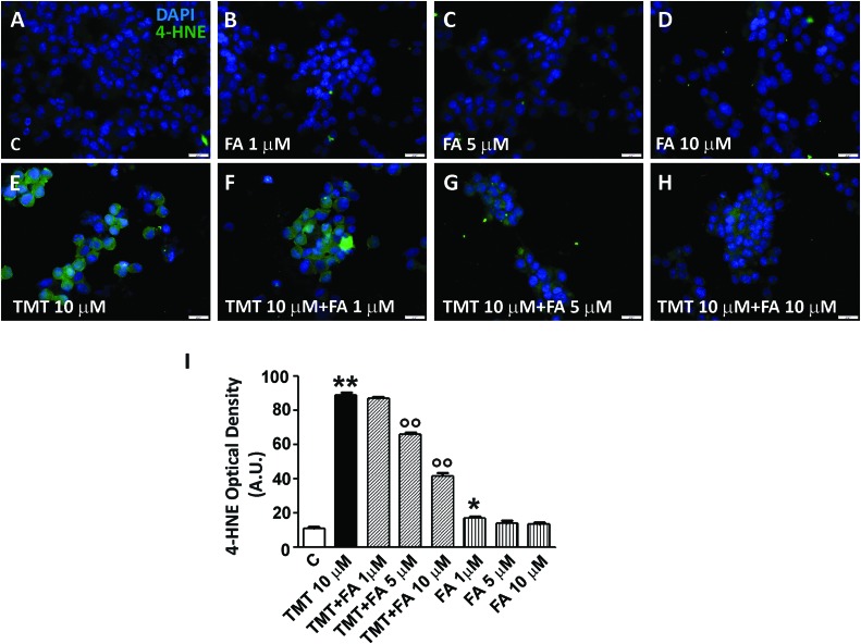
Ferulic acid counteracts the increase of lipid peroxidation induced by TMT in a dose-dependent manner. (A–H) Representative immunofluorescence images of SH-SY5Y cells treated with TMT (10 μM) and/or FA (1–10 μM) and double stained with DAPI (blue) and anti-4-HNE antibody (green). 4-HNE labeling was faint in control cells (A), but mildly increased in cells treated with different doses of FA (B–D). After TMT treatment, there was a marked increase of 4-HNE fluorescence (E) that decreased in a dose-dependent manner when FA was added (F–H). (I) Histograms (mean ± SEM) showing optical density quantification in treated cells for 4-HNE labeling. Data are representative of three independent experiments; each count was performed on five fields randomly selected for each experimental condition. Scale bar (A–H) 20 μm. ∗P < 0.05, ∗∗P < 0.01 vs. controls (C), ∘∘P < 0.01 vs. TMT.

