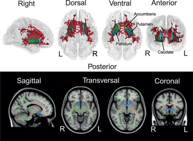
Figure 1.
Reward dependence and WM microstructure. The spatial distribution of voxels showing significant relationships between reward dependence (RD) and FA. Linear effects of RD were tested voxelwise with GLMs while covarying for age and sex. Significant effects (P < 0.0125, fully corrected for multiple comparisons across space and Bonferroni adjusted) are shown as 3D renderings in right, dorsal, ventral, and anterior view on a reconstructed glass cerebrum. Red illustrates voxels showing negative associations with RD and FA (white panel). Striatal structures are illustrated for anatomical reference. The lower (black) panel depicts the same effects superimposed on sagittal, transversal, and coronal slices of a Montreal Neurological Institute (MNI) template brain. Red color indicates significant negative associations between RD and FA. Green areas are the nonsignificant remains of the skeleton. Nucleus accumbens is shown in green for reference. Briefly, associations between RD and FA are predominantly expressed in anterior regions and in the WM surrounding the corpus striatum central to reward processing. Portrayed sections are sagittal (x-coordinate, 103), transversal (z-coordinate, 62 and 76), and coronal (y-coordinate, 142) referring to the MNI coordinate (millimeter) system. R indicates right; L, left.

