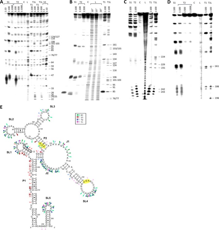
FIGURE 2.
Secondary structure of bsrE mRNA (255 nt). A–D, secondary structure probing of bsrE RNA with RNases as in Fig. 1. Digested RNAs were separated on 8% (A, B, and D) or 15% (C) denaturing gels. A and B, 5′-labeled bsrE; C and D, 3′-labeled bsrE. Autoradiograms are shown. C, L, and T1L are as defined in the legend to Fig. 1. E, proposed secondary structure of bsrE mRNA. A structure inferred from the cleavage data in A–D is depicted. Symbols are as defined in the legend to Fig. 1. The long double-stranded helix P1, helix P2, the single-stranded regions J1 and J2, and the four main stem loops SL1–SL5 are indicated. The bsrE SD sequence is boxed. Start and stop codon are shaded in yellow.

