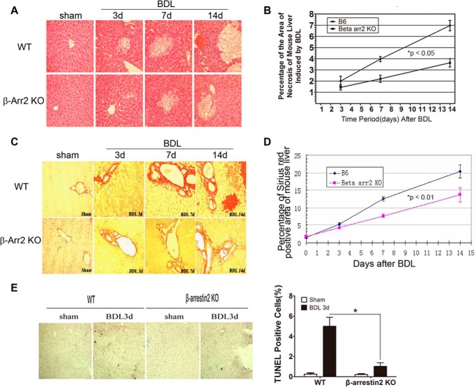
FIGURE 2.
Hepatocyte apoptosis is reduced in Arrb2 KO mice following BDL. Hepatic morphology was determined by H&E staining in WT mice and Arrb2 KO mice following sham operation or BDL at different times (A and B). A, areas of bile infarcts in WT mice are more prominent compared with Arrb2 KO mice (×400). B, morphometric evaluation of the percentage of liver area with bile infarcts in WT mice and Arrb2 KO mice by Image Pro Plus Version 6.0. *, p < 0.05, Arrb2 KO mice versus WT mice. Hepatic morphology was examined by sirius red staining in WT mice and Arrb2 KO mice at different time points after BDL for C and D. C, sirius red staining showed significantly decreased hepatic fibrosis (red) in Arrb2 KO mice compared with WT mice (×400). D, morphometric evaluation of percentage of liver with hepatic fibrosis using Image Pro Plus Version 6.0. *, p < 0.01, Arrb2 KO mice versus WT mice. E, 3 days after BDL in WT and Arrb2 KO mice, the mice were sacrificed to obtain the livers. Apoptotic cells (dark cells) were determined by TUNEL assay. Magnification ×40. The bar graph at right shows the percentage of TUNEL-positive cells. *, p < 0.01.

